Jutapak Jenkitkonchai, Kittiporn Punuch , Teerapat Paringkarn, Teerapat Wannawittayapa, Manop Pithukpakorn, Panitta Sitthinamsuwan, Manasmon Chairatchaneeboon, Varodom Charoensawan
Abstract
Melanoma is one of the most aggressive skin cancers with high mortality rates.1 It frequently exhibits high heterogeneity, with diverse genetic and phenotypic characteristics, which often complicate accurate diagnosis, classification and treatment planning.2 A previous study showed that melanoma tumours with high heterogeneity tend to exhibit reduced infiltration of antitumour immune cells, lower expression of immunomodulatory genes and poorer survival.
Single-cell RNA sequencing has emerged as a powerful technology for investigating melanoma cell heterogeneity and identifying subpopulations with distinct gene expression profiles; however, it lacks spatial resolution and cannot capture the positional context of cells within tissue samples. Alternatively, spatial gene expression technologies enable the direct labelling of hundreds to thousands of transcripts within tissue sections. However, their application to skin tissue remains challenging due to the dense, fibrous dermal stroma and layered architecture of the epidermis, not to mention high intrinsic autofluorescence from melanin and other endogenous fluorophores that may interfere with imaging-based readouts.
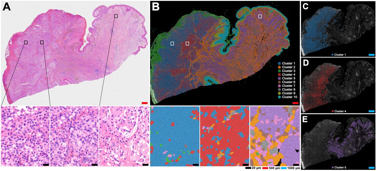
Here, we implement an in situ spatial gene expression method, Xenium (10x Genomics, Pleasanton, CA), to investigate cellular heterogeneity within a congenital melanocytic nevus (CMN). A woman in her 90s with diabetes, hypertension and dyslipidaemia presented with a 3-month history of a progressively enlarging mass on her back. A biopsy confirmed malignant melanoma arising in a CMN (Breslow thickness 3.0 mm, Clark level III, ulcerated, mitotic rate 10–15 per mm²) without angiolymphatic or perineural invasion. Genetic analysis using a multi-target next-generation sequencing panel on a Formalin-Fixed Paraffin-Embedded (FFPE) tissue identified a BRAF mutation. A wide local excision and sentinel node biopsy were performed, showing no residual melanoma and negative sentinel nodes (0/2). CT imaging showed no distant metastasis, and the patient was therefore diagnosed with stage IIB melanoma (pT3bN0M0, AJCC 8th edition). We performed spatial gene expression analysis of the FFPE tissue section (DV200=72.61%) from the patient described above with the predesigned ‘Human Skin’ panel (260 targeted genes) using the Xenium In Situ platform. Additionally, H&E staining was also conducted on the post-Xenium slide (figure 1A). Data were generated from the on-instrument Xenium Onboard Analysis (XOA) pipeline, and we observed a total of 502,626 cells detected and 80,004,559 transcripts (Q score≥20), with a median of 139 transcripts per cell. In the XOA pipeline, before performing downstream analysis, including clustering and differential expression, cells with zero transcript counts were filtered (see full ‘Materials and methods’ section in online supplemental material).